Up to 70% less than Europe.



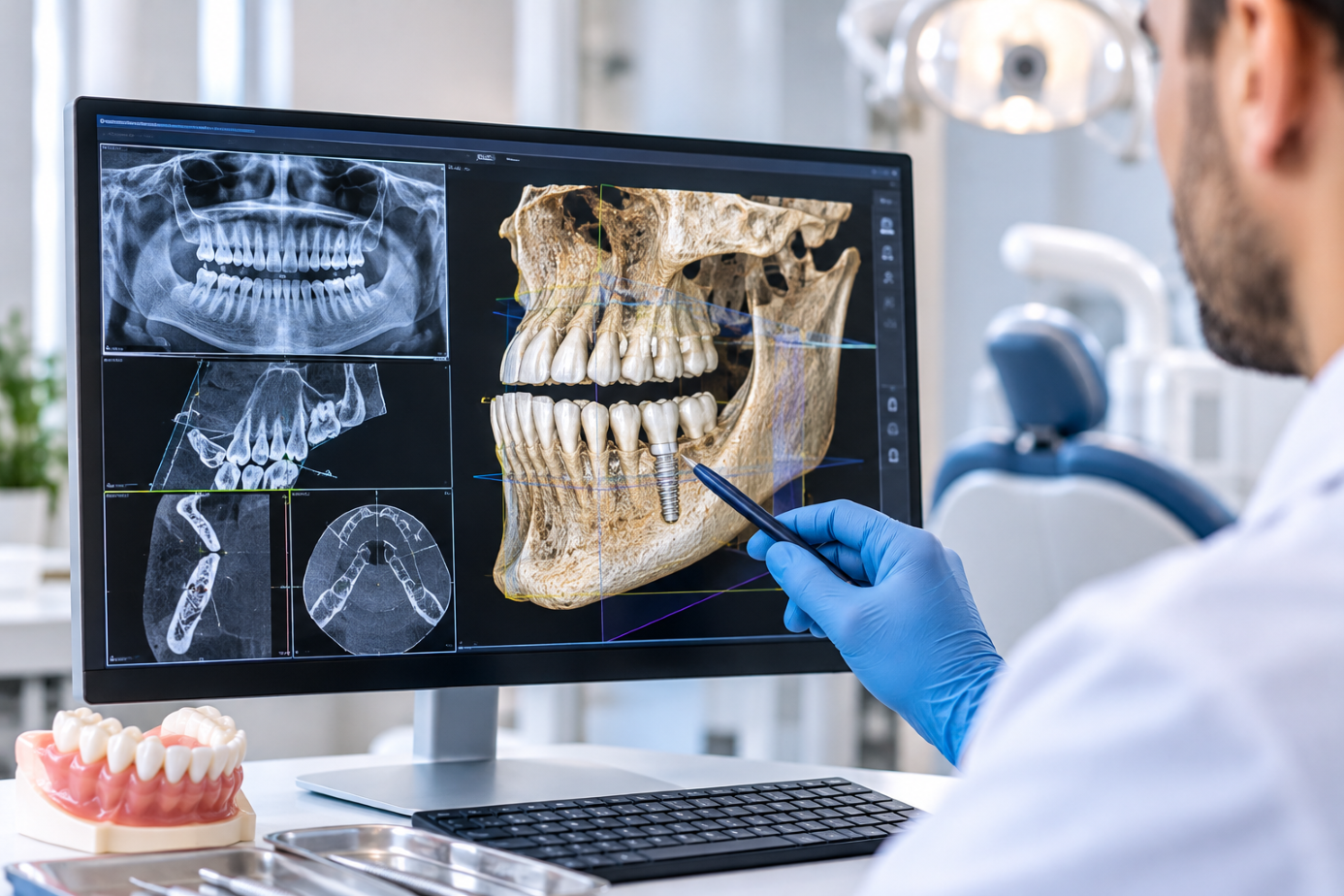
Replacing missing teeth with dental implants requires more than selecting an implant and placing it into the jawbone. Successful treatment depends on accurate diagnosis, detailed anatomical assessment, careful surgical planning, and an understanding of each patient’s individual bone structure.
Modern digital dentistry has transformed the way dentists evaluate implant cases. One of the most valuable diagnostic technologies is Cone Beam Computed Tomography (CBCT), which provides three-dimensional images of the teeth, jawbone, and surrounding anatomical structures.
3D Dental Imaging for Implants allows dentists to examine areas that cannot be fully assessed using conventional two-dimensional dental X-rays. This additional information can help improve treatment planning, identify potential complications, and support safer surgical decisions.
1. What Is 3D Dental Imaging?
Three-dimensional dental imaging is a diagnostic technique that uses specialised scanning equipment to create a three-dimensional representation of the patient’s oral and maxillofacial structures.
One commonly used technology is Cone Beam Computed Tomography, also known as CBCT.
Unlike conventional dental X-rays, which produce flat images, CBCT captures multiple projections that are reconstructed into a three-dimensional dataset. Dentists can then examine the jaw from different angles and evaluate specific anatomical areas.
A CBCT examination may help assess:
- Jawbone height, width, and shape.
- Bone volume available for implant placement.
- The position of nerves and blood vessels.
- The location and dimensions of the maxillary sinuses.
- The relationship between neighbouring teeth and proposed implant sites.
- Existing dental conditions that may affect treatment planning.
CBCT does not automatically need to be performed for every dental implant patient. The dentist should determine whether the additional information is clinically necessary and select an appropriate scanning field of view and exposure settings.
2. Why Is CBCT Important Before Dental Implant Surgery?
Dental implants must be positioned within the available bone while respecting important anatomical structures.
A conventional panoramic X-ray can provide a useful overview of the jaw, but it does not show bone width or depth accurately because it compresses three-dimensional anatomy into a two-dimensional image.
This is where CBCT can provide additional diagnostic information.
Evaluating Bone Height and Width
One of the first considerations in implant planning is whether there is enough bone to accommodate the proposed implant.
The dentist evaluates the available bone dimensions to determine:
- Whether the implant can be placed in the planned position.
- Which implant diameter and length may be appropriate.
- Whether the bone shape creates surgical limitations.
- Whether bone augmentation may be necessary.
Bone volume alone does not determine implant suitability. Bone density, quality, anatomical position, and the patient’s clinical condition must also be considered.
Identifying Important Nerve Structures
In the lower jaw, the inferior alveolar nerve is an important anatomical structure that must be protected during implant surgery.
CBCT can help identify the position of the mandibular canal and estimate the available distance between the planned implant and the nerve.
This information helps the dentist select an appropriate implant position and dimensions while reducing the risk of nerve injury.
Evaluating the Maxillary Sinus
In the upper posterior jaw, the maxillary sinus can limit the available bone height for implant placement.
Three-dimensional imaging helps the dentist evaluate the sinus floor, remaining bone, and anatomical variations.
If insufficient bone is available, procedures such as sinus augmentation may be considered. However, this decision requires an individual clinical assessment rather than relying on imaging measurements alone.
3. How Does a CBCT Scan Work?
A CBCT examination is generally a quick and non-invasive diagnostic procedure.
The process usually involves the following stages:
Step 1: Patient preparation
The patient may be asked to remove metal objects such as earrings, necklaces, or removable dental appliances that could interfere with image quality.
Step 2: Positioning
The patient stands or sits in the scanner according to the equipment design. The dental team positions the head carefully to minimise movement.
Step 3: Image acquisition
The scanner rotates around the patient’s head and captures multiple X-ray projections from different angles.
Step 4: Digital reconstruction
Specialised software processes the images to create a three-dimensional dataset.
Step 5: Clinical evaluation
The dentist examines the relevant anatomical structures and uses the findings alongside the clinical examination and other diagnostic records to develop a treatment plan.
The scan itself is usually completed within a short period. However, the time required to interpret the images and plan surgery depends on the complexity of the case.
4. How Is 3D Imaging Used in Implant Treatment Planning?
The value of three-dimensional imaging goes beyond simply viewing the jawbone. It can help the dentist plan the position, direction, and dimensions of the implant before surgery.
During 3D Dental Imaging for Implants, the dentist can assess the proposed implant position in relation to the available bone and surrounding structures.
The planning process may include:
Implant Position Assessment
The dentist evaluates where the implant should be placed to provide appropriate support for the future crown or bridge.
Implant positioning should consider both surgical anatomy and the final restoration. An implant that is positioned only according to available bone may create difficulties when designing the final tooth.
Implant Angulation
The angle of an implant affects how the final restoration will emerge from the gum and connect to the supporting components.
Digital planning can help assess whether the proposed angle is compatible with the desired crown position, bite, and surrounding anatomy.
Implant Dimensions
The dentist considers the appropriate implant length and diameter based on the available bone and the restorative requirements.
Larger implants are not automatically better. The dimensions must be selected according to the patient’s anatomy and the manufacturer’s clinical recommendations.
Surgical Guide Planning
In selected cases, CBCT data can be combined with digital dental scans to create a virtual implant plan.
This information may be used to design a computer-guided surgical template. Such guides can help transfer the planned implant position to the surgical procedure.
Guided surgery can improve the accuracy of implant positioning in suitable cases, but it does not eliminate surgical risks or replace the dentist’s clinical judgement.
5. Can 3D Imaging Identify the Need for Bone Grafting?
One of the important advantages of CBCT is its ability to evaluate the dimensions and shape of the jawbone.
After tooth extraction, the surrounding bone may gradually shrink. Some patients may therefore have insufficient bone volume for conventional implant placement.
Depending on the findings, the dentist may consider:
- Guided bone regeneration.
- Localised bone grafting.
- Sinus augmentation in the upper posterior jaw.
- Alternative implant positions or dimensions.
- Different restorative treatment options.
Not every patient with reduced bone volume requires grafting. In certain cases, alternative implant designs or treatment approaches may be appropriate.
The final decision depends on the anatomical findings, clinical examination, and the intended restoration.
6. Does 3D Dental Imaging Reduce Implant Surgery Risks?
Three-dimensional imaging can help identify anatomical challenges before surgery, allowing the dentist to plan around important structures.
However, it is important to understand that imaging does not guarantee a complication-free procedure.
Potential complications associated with implant surgery include:
- Infection or delayed healing.
- Damage to nearby nerves or anatomical structures.
- Sinus-related complications in upper jaw surgery.
- Inadequate initial implant stability.
- Failure of osseointegration.
- Bone loss or inflammation around the implant.
- Mechanical complications affecting implant components.
CBCT can help assess certain anatomical risks, but it cannot predict every biological or surgical outcome.
The dentist must also consider the patient’s medical history, smoking habits, gum health, oral hygiene, medications, and general healing capacity.
7. Is CBCT Safe? Understanding Radiation Exposure
CBCT uses ionising radiation, so its use should always be based on clinical necessity.
Although dental CBCT examinations generally use lower radiation doses than conventional medical CT examinations, the actual dose varies considerably depending on the machine, field of view, exposure settings, and scanning protocol.
The dental team should:
- Request CBCT only when it is expected to provide useful diagnostic information.
- Use the smallest appropriate field of view.
- Select exposure settings suitable for the clinical purpose.
- Avoid unnecessary repeat examinations.
- Consider previous imaging when available.
Patients should inform the dental team if they are pregnant or may be pregnant before undergoing the examination.
The benefits of additional diagnostic information should always be balanced against radiation exposure.
8. What Happens After the CBCT Scan?
Once the scan has been completed, the dentist reviews the images and combines the findings with the clinical examination.
The patient should receive an explanation of the proposed treatment, including any anatomical limitations or additional procedures that may be required.
The next steps may involve:
- Confirmation of the proposed implant positions.
- Selection of suitable implant dimensions.
- Discussion of bone grafting or sinus augmentation if needed.
- Preparation of the surgical and restorative plan.
- Explanation of treatment stages, costs, and expected healing.
- Scheduling of surgery and follow-up appointments.
A scan is a diagnostic tool, not a treatment plan by itself. The dentist must interpret the findings and explain how they affect the individual patient’s treatment.
9. Does 3D Imaging Make Dental Implant Treatment More Expensive?
The cost of implant treatment depends on several clinical and technical factors.
A CBCT scan may be included in a dental implant package or charged separately, depending on the clinic’s pricing policy.
Other factors that may influence the total cost include:
- Number of implants required.
- Implant brand and system.
- Bone grafting or sinus augmentation.
- Surgical guide production, if indicated.
- Temporary and final restorations.
- Laboratory procedures.
- Number of treatment visits.
Patients should request a written quotation explaining which diagnostic procedures are included and whether additional surgical requirements could change the original estimate.
It is important not to choose a clinic simply because it offers the cheapest scan or implant package. Diagnostic quality, appropriate treatment planning, surgical experience, and long-term aftercare should all be considered.
10. The Patient Journey: Dental Implant Planning in Izmir
For international patients, understanding the treatment journey before travelling is especially important.
At Optimum Dental Turkey, the process begins with an initial assessment of the patient’s dental concerns and available records.
Before arrival: Patients can share photographs and previous dental X-rays where available. These provide preliminary information but cannot replace a clinical examination.
During the first appointment: The dentist examines the mouth and determines which diagnostic records are necessary. If three-dimensional imaging is clinically indicated and available, the scan can be used to assess the jawbone and relevant anatomical structures.
Treatment planning: The dentist discusses the implant positions, possible additional procedures, treatment alternatives, estimated costs, and expected healing period.
Surgical treatment: Implant placement is carried out according to the agreed clinical plan. Temporary restorations may be provided when appropriate.
Healing and follow-up: The patient receives post-operative instructions and information about the healing period. If a second visit is needed for the final restoration, the expected timing is discussed before the patient returns home.
Patients looking for Dental treatment in Izmir should also consider the clinic’s communication, diagnostic facilities, and aftercare arrangements.
Optimum Dental Turkey provides patient support and VIP transfers between the airport, hotel, and clinic. The clinic is conveniently located for visitors travelling through Izmir Adnan Menderes Airport, near Izmir airport. Accommodation and transfer arrangements should be confirmed as part of the individual treatment package.
11. Frequently Asked Questions About 3D Dental Imaging
Is a CBCT scan necessary for every dental implant?
No. The dentist determines whether CBCT is clinically justified. In some straightforward cases, conventional imaging and clinical examination may provide sufficient information. Three-dimensional imaging is particularly useful when additional anatomical detail could influence treatment decisions.
How long does a CBCT scan take?
The actual image acquisition usually takes less than a minute, although preparation and positioning may take additional time. Reviewing and interpreting the scan requires a separate clinical assessment.
Can CBCT show if I have enough bone for dental implants?
CBCT can provide measurements of bone height, width, and shape. However, the dentist must also consider bone quality, clinical findings, implant positioning, and restorative requirements.
Can a CBCT scan detect nerve problems?
CBCT can show the location and course of certain important nerve pathways, such as the mandibular canal. It does not directly diagnose every nerve condition.
Is guided implant surgery always more accurate?
Guided surgery can help transfer a digital plan to the surgical procedure in selected cases. Accuracy depends on scan quality, planning, guide design, guide stability, and surgical technique. It is not automatically necessary for every patient.
Can I receive my implant on the same day as my scan?
A CBCT scan can support treatment planning, but same-day implant placement depends on the clinical assessment, surgical preparation, and availability of the required facilities. It should never be assumed before the dentist has confirmed suitability.
Conclusion: Better Planning Starts with Better Diagnosis
Modern dental implant treatment relies on accurate diagnosis and careful planning. CBCT technology gives dentists a more detailed understanding of the jawbone and surrounding anatomical structures, helping them evaluate potential challenges before surgery.
3D Dental Imaging for Implants can be particularly valuable when assessing bone availability, identifying important anatomical structures, planning implant dimensions, and considering additional procedures.
At Optimum Dental Turkey, diagnostic decisions should be based on individual clinical needs rather than routine scanning. The goal is to develop a treatment plan that considers safety, function, natural tooth preservation, and long-term oral health.
Optimum Dental Turkey Advantages

Free Consultation
Contact Us for an Online or In-Person Consultation
During your consultation, you will receive:
✔️ A comprehensive dental examination
✔️ A personalized treatment plan
✔️ A full cost estimate and treatment strategy

Customer Care Manager
By communicating with our professional translators, we ensure that everyone receives health care comfortably.


VIP Services
Reach our clinic with luxury vehicles with our VIP Service and receive accommodation in luxury hotels.